Skip to content Skip to sidebar Skip to footer

Retrocardiac Air Space Disease

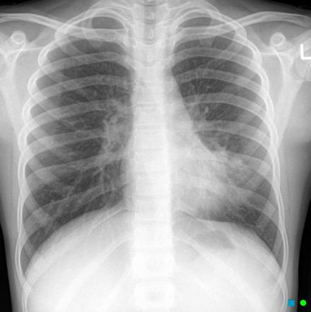
Patchy Airspace Opacification In The Left Retrocardiac Region With Download Scientific Diagram

Patchy Airspace Opacification In The Left Retrocardiac Region With Download Scientific Diagram

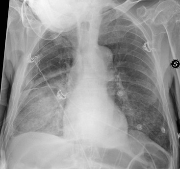
Retrocardiac air space disease. Airspace disease is considered chronic when it persists beyond 4-6 weeks after treatment. Tendency to coalesce near hila. The margins of airspace disease are fuzzy and indistinct.

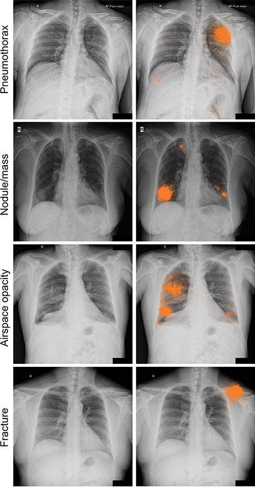
Air-space opacification is a descriptive term that refers to filling of the lung parenchyma with material that attenuates x-rays more than the unaffected surrounding lung tissue. Consolidation or ground-glass opacity occurs when alveolar air is replaced by fluid pus blood cells or other material. Test results for infl uenza virus respiratory fl uorescent antibodies D3 Ultra DFA Respiratory Virus Screening and In-fectious Disease Kit.

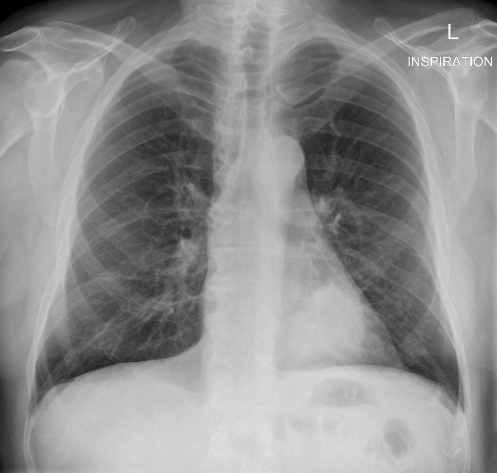
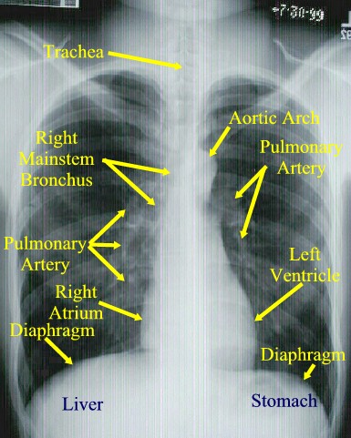
Air cant get through the consolidation so your lung cant do its job of bringing in fresh air and removing the air your body has used. The location retrocardiac - behind the heart shouldnt make a difference. Identifying multifocal air-space disease on CXR can be a significant clue to COVID-19 pneumonia.

By early detection and proper evaluation of the abnormal retrocardiac shadows the radiologist may establish the diagnosis before the clinical signs and symptoms of compression of large blood vessels and nerves become. Retrocardiac Air Space Disease. Air space opacification is a descriptive term that refers to filling of the pulmonary tree with material that attenuates x-rays more than the surrounding lung parenchyma.

This unusual type of bibasilar atelectasis happens when the lung is trapped as a result of pleural disease while being devoid of air. The opacities tend to be confluent merging into one another. These represent air-space or acinar nodules.

Patchy diffuse air space consolidation. Diagnostic Hy-brids Inc Athens OH USA and virus culture were negative. There are a number of different causes of insult to the alveoli including build up of fluid hemorrhage infection malignancy and build up of protein and mineral deposits.

It is one of the many patterns of lung opacification and is equivalent to the pathological diagnosis of pulmonary consolidation. The latter will often reveal air trapping on expiratory images 25-27.

Air Space Opacification Radiology Reference Article Radiopaedia Org

Air Space Opacification Radiology Reference Article Radiopaedia Org

Https Vicenteayalabermeo Files Wordpress Com 2011 07 Dr Juan 2 Pdf

Https Vicenteayalabermeo Files Wordpress Com 2011 07 Dr Juan 2 Pdf

Retrosternal Airspace Radiology Reference Article Radiopaedia Org

Retrosternal Airspace Radiology Reference Article Radiopaedia Org

Dr Pepe S Diploma Casebook Case 115 Solved Blog

Dr Pepe S Diploma Casebook Case 115 Solved Blog

Lateral Chest X Ray Demonstrated Increase In Retrosternal And Download Scientific Diagram

Lateral Chest X Ray Demonstrated Increase In Retrosternal And Download Scientific Diagram

Chronic Airspace Diseases Sciencedirect

Chronic Airspace Diseases Sciencedirect

3 Basic Patterns In Lung Disease Radiology Key

3 Basic Patterns In Lung Disease Radiology Key

Http Pdf Posterng Netkey At Download Index Php Module Get Pdf By Id Poster Id 40277

Http Pdf Posterng Netkey At Download Index Php Module Get Pdf By Id Poster Id 40277

Chest Imaging

Chest Imaging

Chest X Ray Showed Consolidation On Left Lower Lung Fields Especially Download Scientific Diagram

Chest X Ray Showed Consolidation On Left Lower Lung Fields Especially Download Scientific Diagram

A Pain In The Neck Nejm

A Pain In The Neck Nejm

Serial Chest X Rays Showing Diffuse Airspace Consolidation Involving Download Scientific Diagram

Serial Chest X Rays Showing Diffuse Airspace Consolidation Involving Download Scientific Diagram

Http Unofficialguidetomedicine Com Wp Content Uploads 2017 01 Ugrad Chest Preview Pdf

Http Unofficialguidetomedicine Com Wp Content Uploads 2017 01 Ugrad Chest Preview Pdf

Http Unofficialguidetomedicine Com Wp Content Uploads 2017 01 Ugrad Chest Preview Pdf

Http Unofficialguidetomedicine Com Wp Content Uploads 2017 01 Ugrad Chest Preview Pdf

Hemidiaphragm An Overview Sciencedirect Topics

Hemidiaphragm An Overview Sciencedirect Topics

Atraumatic Conditions Of The Chest Anesthesia Key

Atraumatic Conditions Of The Chest Anesthesia Key

Review Areas On Chest Radiograph Radiology Reference Article Radiopaedia Org

Review Areas On Chest Radiograph Radiology Reference Article Radiopaedia Org

Radiologia Brasileira Achados Iniciais Na Radiografia De Torax Em Uma Populacao Pediatrica Com Diagnostico De Infeccao Viral Por H1n1

Radiologia Brasileira Achados Iniciais Na Radiografia De Torax Em Uma Populacao Pediatrica Com Diagnostico De Infeccao Viral Por H1n1

X Ray Atlas Chest X Ray Glowm

X Ray Atlas Chest X Ray Glowm

Esophageal Dilatation With Air Esophogram Sign Achalasia Images Diagnosis Treatment Options Answer Review Thoracic Imaging

Esophageal Dilatation With Air Esophogram Sign Achalasia Images Diagnosis Treatment Options Answer Review Thoracic Imaging

Acute Chest Syndrome And Covid 19 In Sickle Cell Disease Pediatric Patients

Acute Chest Syndrome And Covid 19 In Sickle Cell Disease Pediatric Patients

Chest X Ray Interpretation And Teaching

Chest X Ray Interpretation And Teaching

Atraumatic Conditions Of The Chest Anesthesia Key

Atraumatic Conditions Of The Chest Anesthesia Key

Https Encrypted Tbn0 Gstatic Com Images Q Tbn And9gcrlfdftzzajbjuisei P649ftmnnnmumpqvqvh5hb7gkztdnkd6 Usqp Cau

Https Encrypted Tbn0 Gstatic Com Images Q Tbn And9gcrlfdftzzajbjuisei P649ftmnnnmumpqvqvh5hb7gkztdnkd6 Usqp Cau

Radiologia Brasileira Achados Iniciais Na Radiografia De Torax Em Uma Populacao Pediatrica Com Diagnostico De Infeccao Viral Por H1n1

Radiologia Brasileira Achados Iniciais Na Radiografia De Torax Em Uma Populacao Pediatrica Com Diagnostico De Infeccao Viral Por H1n1

Night Float Module Interpretation Of Chest Radiographs Ppt Video Online Download

Night Float Module Interpretation Of Chest Radiographs Ppt Video Online Download

Imaging Manifestations Of Blastomycosis A Pulmonary Infection With Potential Dissemination Radiographics

Imaging Manifestations Of Blastomycosis A Pulmonary Infection With Potential Dissemination Radiographics

Chronic Airspace Diseases Sciencedirect

Chronic Airspace Diseases Sciencedirect

Airspace Diseases Radiology Key

Airspace Diseases Radiology Key

Left Lower Lobe Retrocardiac Lung Cancer Radiology Case Radiopaedia Org

Left Lower Lobe Retrocardiac Lung Cancer Radiology Case Radiopaedia Org

Chest European Diploma Of Radiology

Chest European Diploma Of Radiology

Epos

Epos

Https Link Springer Com Content Pdf 10 1007 2f0 306 46834 4 6 Pdf

Https Link Springer Com Content Pdf 10 1007 2f0 306 46834 4 6 Pdf

Chest Xxxx And Lateral Open I

Chest Xxxx And Lateral Open I

Achalasia Images Diagnosis Treatment Options Answer Review Thoracic Imaging

Achalasia Images Diagnosis Treatment Options Answer Review Thoracic Imaging

Deep Learning For Chest Radiograph Diagnosis A Retrospective Comparison Of The Chexnext Algorithm To Practicing Radiologists

Deep Learning For Chest Radiograph Diagnosis A Retrospective Comparison Of The Chexnext Algorithm To Practicing Radiologists

Imaging Evaluation Of Covid 19 In The Emergency Department Abstract Europe Pmc

Imaging Evaluation Of Covid 19 In The Emergency Department Abstract Europe Pmc

Enews Exclusive Radiology Today Magazine

Enews Exclusive Radiology Today Magazine

Http Pdf Posterng Netkey At Download Index Php Module Get Pdf By Id Poster Id 40277

Http Pdf Posterng Netkey At Download Index Php Module Get Pdf By Id Poster Id 40277

Presentation Materials Basic Chest Radiology For The Tb

Presentation Materials Basic Chest Radiology For The Tb

Resorptive Atelectasis Of Lul Cancer Lung

Resorptive Atelectasis Of Lul Cancer Lung

Ring Of Fire Appearance In Covid 19 Pneumonia Bmj Case Reports

Ring Of Fire Appearance In Covid 19 Pneumonia Bmj Case Reports

Chest X Ray

Chest X Ray

Https Www Bir Org Uk Media 258608 Mark Rodriguez Philips Trainee For Excellence Unofficial Guide To Radiology Pdf

Https Www Bir Org Uk Media 258608 Mark Rodriguez Philips Trainee For Excellence Unofficial Guide To Radiology Pdf

Hiatal Hernia Explanation Of Chest X Ray Findings Youtube

Hiatal Hernia Explanation Of Chest X Ray Findings Youtube

Cardiothoracic Imaging Radiology Key

Cardiothoracic Imaging Radiology Key

Pulmonary Fibrosis On The Lateral Chest Radiograph Kerley D Lines Revisited Insights Into Imaging Full Text

Pulmonary Fibrosis On The Lateral Chest Radiograph Kerley D Lines Revisited Insights Into Imaging Full Text

Pneumonia Springerlink

Pneumonia Springerlink

Https Encrypted Tbn0 Gstatic Com Images Q Tbn And9gcqnvm3zlkgdzb2bsnt1cyl1a Tkeegf Ie0unitga7cn0ww4wd9 Usqp Cau

Https Encrypted Tbn0 Gstatic Com Images Q Tbn And9gcqnvm3zlkgdzb2bsnt1cyl1a Tkeegf Ie0unitga7cn0ww4wd9 Usqp Cau

Alveolar lung diseases are classified as processes that affect these units that ultimately lead to issues with ventilation.

Airspace disease is considered chronic when it persists beyond 4-6 weeks after treatment. A single retrocardiac air-fluid level on a chest radiograph typically implies the presence of a sliding hiatal hernia. This unusual type of bibasilar atelectasis happens when the lung is trapped as a result of pleural disease while being devoid of air. Airspace disease also known as alveolar lung disease is a generic term thats used to describe abnormalities on chest x-ray or CT. The most common clinical causes of this CT pattern of disease include hypersensitivity pneumonitis sarcoidosis atypical infections eg those caused by Mycoplasma pneumoniae with associated bronchiolitis and acute interstitial pneumonia 25. The 2021 edition of ICD-10-CM J984 became effective on October 1 2020. J984 is a billablespecific ICD-10-CM code that can be used to indicate a diagnosis for reimbursement purposes. And retrocardiac air space disease. Retrocardiac Air Space Disease.


The location retrocardiac - behind the heart shouldnt make a difference. It is the radiological correlate of the pathological diagnosis of pulmonary consolidation. The most common clinical causes of this CT pattern of disease include hypersensitivity pneumonitis sarcoidosis atypical infections eg those caused by Mycoplasma pneumoniae with associated bronchiolitis and acute interstitial pneumonia 25. The particular way the lung collapses can often produce a. Acinar or air-space nodules. CT 5-mm slice thickness in a patient with bilateral consolidation. Tendency to coalesce near hila.

Post a Comment for "Retrocardiac Air Space Disease"